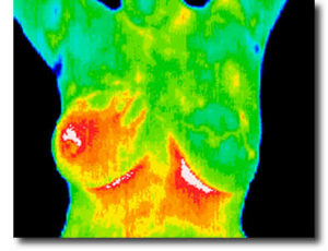
Thermal Breast Scans
 When the time seems right to screen your breasts, we like to encourage women to utilize a technology that is finally coming of age–thermal imaging.
I first learned about thermal imaging in the early 90s, and then it disappeared altogether because of poor protocols, unsystematic training, and inconsistent standards for reporting. But as the technology has improved and standards are becoming more strict, it is once again back on the scene and growing in availability. Care, however, must still be taken in who is performing the test and who is reading the reports.
Properly conducted thermal imaging has been found to identify signs of breast cancer up to 10 years before any other modality, including mammography. Now you can identify cancer signs earlier than previously thought possible. What this means is that you can detect changes in breast tissue that suggests pathology and begin to make the necessary changes to avoid more invasive and aggressive procedures or medical actions. You can also monitor the success of the treatment methods you have chosen. This can help allay many fears, especially for women who choose non-conventional treatment modalities.
The good news for women is that the screening involves no pain, no contact, no compression and most importantly no radiation. Recently I heard a doctor from Hoag Hospital say that thermal imaging is the wave of the future for breast health monitoring. Today, with a quality thermal imaging center, you can have highly-trained medical doctors reading the images with the aid of sophisticated computer programs, elevating the quality of the findings report.
Thermal imaging is for adult women of any age. With two scans, you will have your “breast print” which does not change form scan to scan unless there are changes in pathology. This gives a constant reference point for monitoring your breast health for any condition of concern.
For more information about the thermal imaging center we recommend locally, visit Thermal Imaging of Southern California. This center utilizes top-quality DIDI cameras and sends the reports to Duke University to be read by experienced oncologists and medical doctors. Wherever you choose to have thermal imaging performed, we currently encourage centers that utilize the Duke University reporting because of the quality of staff readers and consistency in training.
